Tech
New tech allows parents to hold a 3D printed version of their unborn baby

US surgeons have developed new 3D printing technology to assist with procedures to correct birth defects in the womb
One hospital in Florida is working with 3D printing developers to create new technology to revolutionise the future of pre-natal care. It aims to make fetal surgery more effective and safer for both the mother and unborn child. The technology allows surgeons to create a detailed 3D rendering of the unborn child to study before surgery.
Surgeons use MRI ultrasound imaging and the 3D printing tech to reduce potential risks in procedures. The hospital says the technology also allows surgeons to plan procedures ahead of time and plan for anticipated obstacles.

The technology could prevent the unborn child from developing neurological disabilities, such as an inability to walk
The hospital is trialling using the tech to plan for in-utero surgery to repair spinal defects. The defects can lead to neurological disabilities, such as being unable to walk.
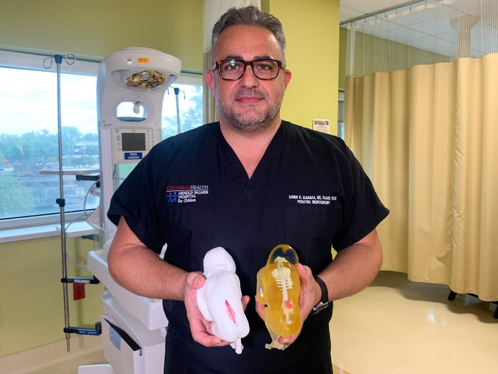
Samer Elbabaa, MD is the medical director of paediatric neurosurgery at Orlando Health, the hospital that developed the technology. She says the tech is “extremely valuable” in cases where defects can’t actually be seen ahead of surgery.

Helping surgeons to identify and analyse potential issues before the baby is even born
“The 3D reconstruction of the fetus can really educate the surgeon on the real-life shape, size and location of the spinal lesion,” she said.
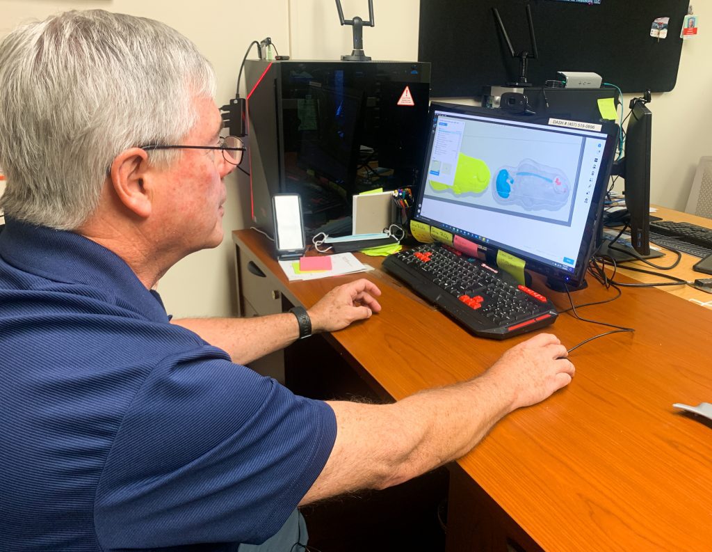
DASH President and CEO Jack Stubbs says the the fetal models will help surgeons plan for procedures. It will also help “reduce the duration of the surgery to limit the developing baby’s exposure,” he said.
“It’s a level of detail that we are not able to see in traditional imaging”
The technology works by enhancing MRI and ultrasound images taken throughout the pregnancy with more accurate 3D details.
These images are then printed as a high-res model using multiple colours and materials. This also allows surgeons to see extra details like skeletal structure, nerves, veins and other issues indicative of spinal abnormalities.
“We are able to create models that are extremely realistic,” said Stubbs. They do this by using a stack of two-dimensional images taken throughout the pregnancy and enhancing them to reconstruct a more accurate visualisation of the fetus.

The 3D-printed models will give both surgeons and parents a clearer picture for what to expect during fetal surgery
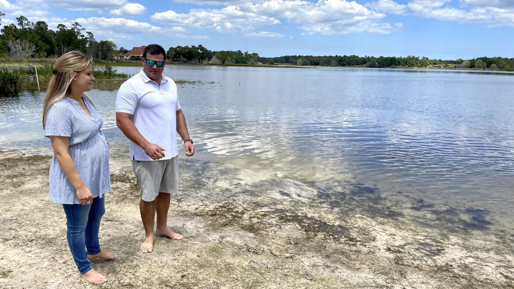
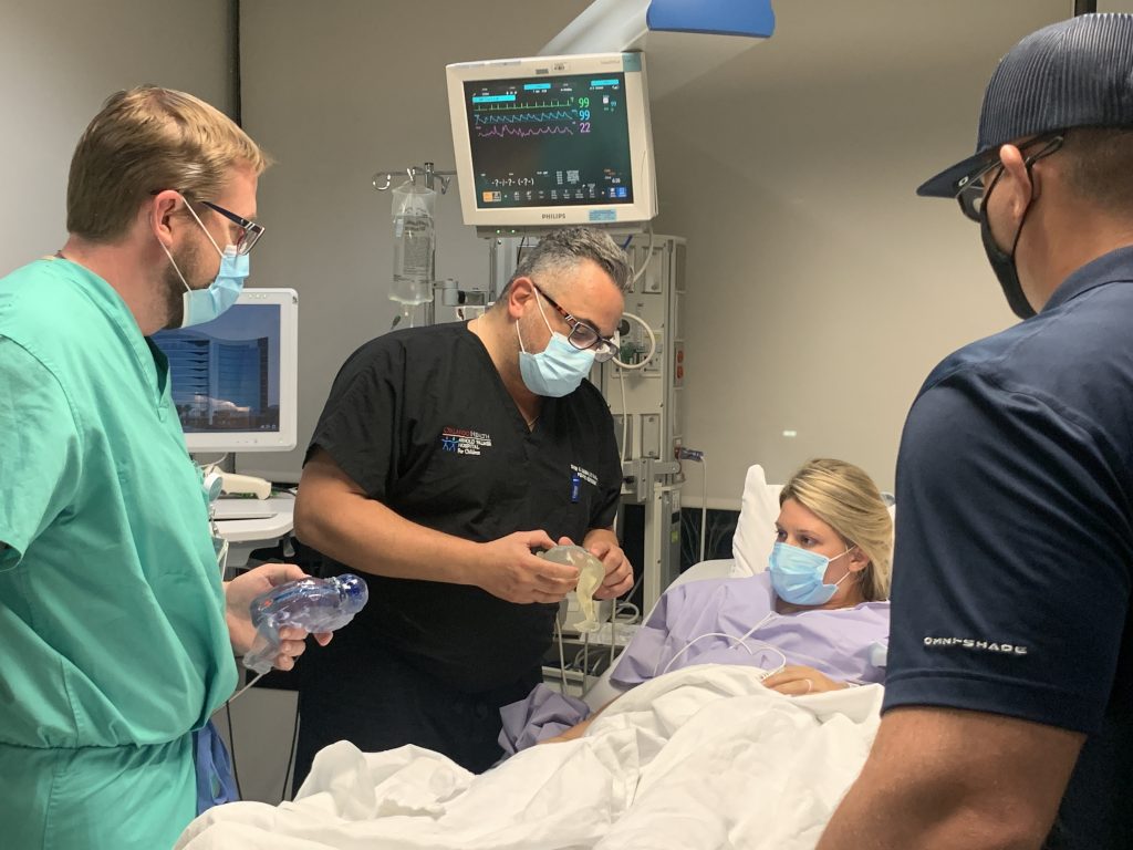
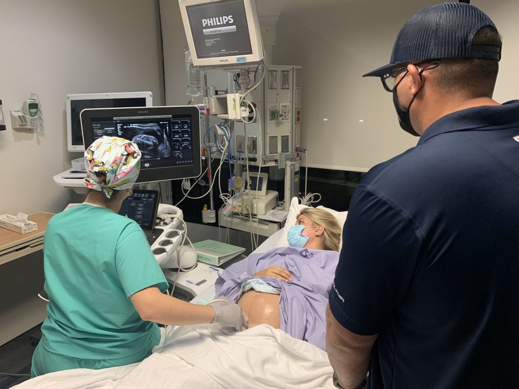
The technology will also help surgeons explain the baby’s condition and potential treatment options to parents. For first-time parents Jared and Jocelyn Rodriguez, it made them more confident about moving forward with surgery.
“We could see the brain and the spine. I looked down at it and thought, ‘I’m holding my daughter right now? That’s pretty awesome.’”
The Rodriguezes say they’re glad this technological development will help give their future daughter the possibility of a healthier future.